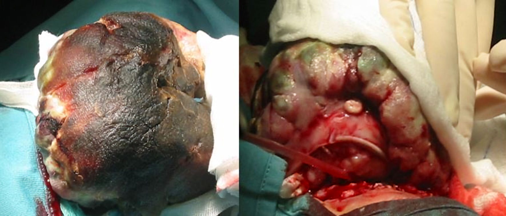
Figure 1. Preoperative pictures. The mass size is comparable to a child’s head.
| International Journal of Clinical Pediatrics, ISSN 1927-1255 print, 1927-1263 online, Open Access |
| Article copyright, the authors; Journal compilation copyright, Int J Clin Pediatr and Elmer Press Inc |
| Journal website https://www.theijcp.org |
Case Report
Volume 13, Number 2, June 2024, pages 54-59
A Five Hundred Six-Gram Giant Orbital Retinoblastoma: An Unusual Case Report
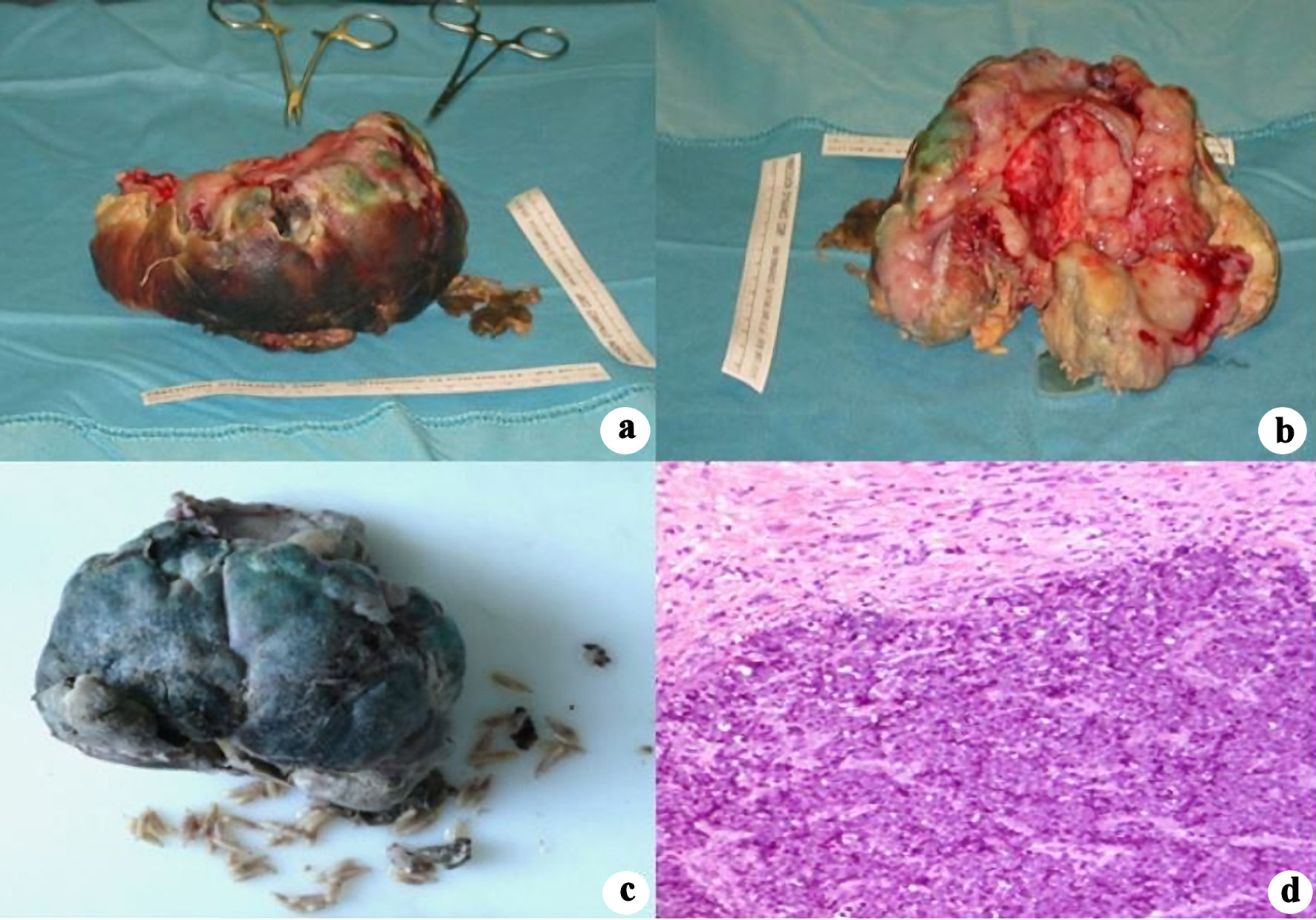
Figures