Figures
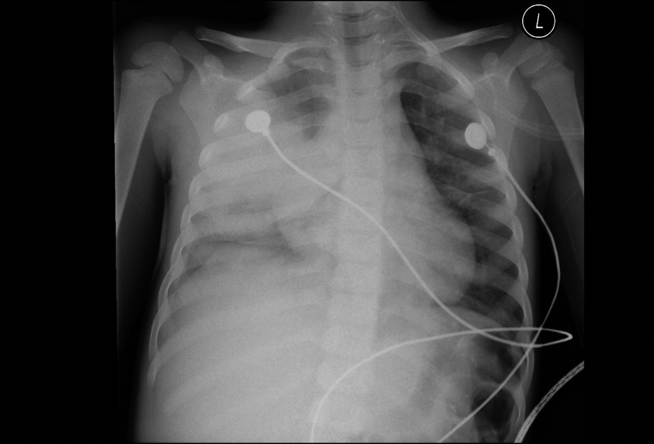
Figure 1. 42-month-old male with acute chest syndrome. Frontal view of the chest reveal consolidations in the right lower lobe, right upper lobe, and left lower lobe. Right-sided pleural effusion is present. Patient was hospitalized for 14 days in the intensive care unit. During hospitalization patient was intubated and his category of clinical course severity was 1. There are consolidations in three lobes with right-sided pleural effusion and his cut-off level in interpreting the chest radiograph was classified at level 1.
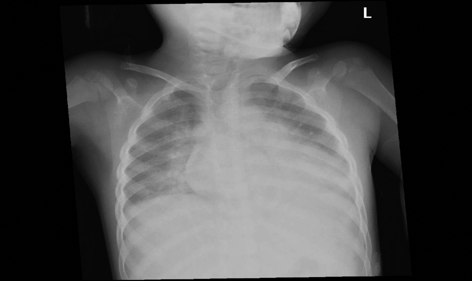
Figure 2. 54-month-old female with acute chest syndrome. Frontal view of the chest reveal consolidation in the right upper lobe, right lower lobe, left lower lobe, and left upper lobe. Patient was hospitalized for 14 days and was intubated in the intensive care unit. His category of clinical severity was 1 and the cut-off level in interpreting the chest radiograph was 2.
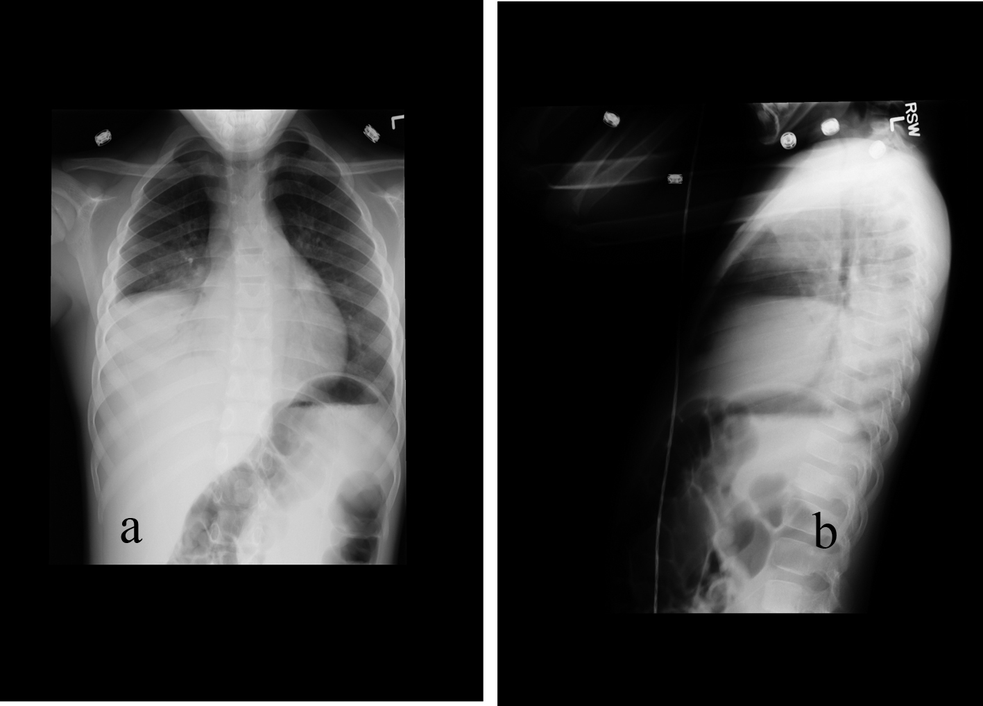
Figure 3. 77-month-old male with acute chest syndrome. Frontal (a) and lateral (b) views of the chest reveal consolidations in the right lower lobe, right middle lobe, and in part of the right upper lobe. Patient was hospitalized for 4 days and had episodes of hypoxia. He was classified in category 2 of severity of clinical course. His cut-off level in interpreting of the chest radiograph was classified at level 3.
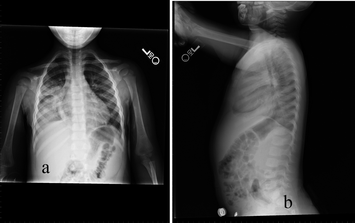
Figure 4. 32-month-old male with acute chest syndrome. Frontal (a) and lateral (b) radiographs of the chest reveal consolidations in the right lower lobe and in the right upper lobe. The patient was hospitalized for 6 days in the intensive care unit and was classified in the clinical course severity as category 3. As there is consolidation in only two lobes the cut-off level for interpreting the chest radiograph was classified at level 4.
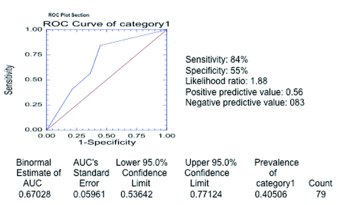
Figure 5. Receiver operating curve in category 1 of severity. The curve was generated with the different threshold levels as defined in the methods section in the entire group of 79 patients. The estimated area under the curve is 0.67028 (above 0.5) confirming that chest radiograph is a valid test to predict severe outcome as defined in category 1.
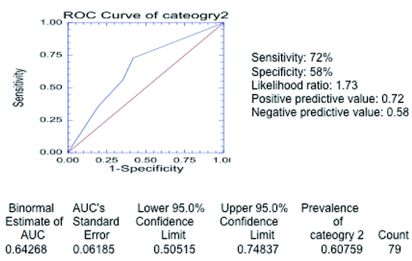
Figure 6. Receiver operating curve in category 2 of severity. The curve was generated with the different threshold levels as defined in the methods section in the entire group of 79 patients. The estimated area under the curve is 0.64268 (above 0.5) confirming that chest radiograph is a valid test to predict severe outcome as defined in category 2.
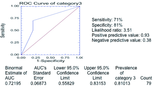
Figure 7. Receiver operating curve in category 3 of severity. The curve was generated with the different threshold levels as defined in the methods section in the entire group of 79 patients. The estimated area under the curve is 0.72195 (above 0.5) confirming that chest radiograph is a valid test to predict severe outcome as defined in category 3.
Tables
Table 1. Distribution of the 17 Patients, Being Below 3 Years Old
| Test + | Test - |
|---|
| test+ means multilobar infiltrates; test- means single lobe infiltrate; severity+ means severe clinical course as defined in category 1; severity- means patients who did not meet the criteria for severe course of category 1. |
| Severity + | 5 | 0 |
| Severity - | 7 | 5 |
Table 2. Distribution of the 24 Patients, Being Between 3 and 6 Years Old
| Test + | Test - |
|---|
| test+ means multilobar infiltrates; test- means single lobe infiltrate; severity+ means severe clinical course as defined in category 1; severity- means patients who did not meet the criteria for severe course of category 1. |
| Severity + | 7 | 2 |
| Severity - | 9 | 6 |
Table 3. Distribution of the 38 Patients, Being Above Years Old
| Test + | Test - |
|---|
| test+ means multilobar infiltrates; test- means single lobe infiltrate; severity+ means severe clinical course as defined in category 1; severity- means patients who did not meet the criteria for severe course of category 1. |
| Severity + | 15 | 3 |
| Severity - | 5 | 15 |
Table 4. Distribution of Our Entire Group of 79 Patients at all Ages in the Category 1 of Severity
| Test + | Test - |
|---|
| test+ means multilobar infiltrates; test- means single lobe infiltrate; severity+ means severe clinical course as defined in category 1; severity- means patients who did not meet the criteria for severe course of category 1. |
| Severity + | 27 | 5 |
| Severity - | 21 | 26 |
Table 5. Distribution of the 17 Patients, Being Below 3 Years Old
| Test + | Test - |
|---|
| test+ means multilobar infiltrates; test- means single lobe infiltrate; severity+ means severe clinical course as defined in category 2; severity- means patients who did not meet the criteria for severe course of category 2. |
| Severity + | 6 | 1 |
| Severity - | 6 | 4 |
Table 6. Distribution of the 24 Patients, Being Between 3 and 6 Years Old
| Test + | Test - |
|---|
| test+ means multilobar infiltrates; test- means single lobe infiltrate; severity+ means severe clinical course as defined in category 2; severity- means patients who did not meet the criteria for severe course of category 2. |
| Severity + | 13 | 6 |
| Severity - | 3 | 2 |
Table 7. Distribution of the 38 Patients, Being Above Years Old
| Test + | Test - |
|---|
| test+ means multilobar infiltrates; test- means single lobe infiltrate; severity+ means severe clinical course as defined in category 2; severity- means patients who did not meet the criteria for severe course of category 2. |
| Severity + | 16 | 6 |
| Severity - | 4 | 12 |
Table 8. Distribution of Our Entire Group of 79 Patients at all Ages in the Category 2 of Severity
| Test + | Test - |
|---|
| test+ means multilobar infiltrates; test- means single lobe infiltrate; severity+ means severe clinical course as defined in category 2; severity- means patients who did not meet the criteria for severe course of category 2. |
| Severity + | 35 | 13 |
| Severity - | 13 | 18 |
Table 9. Distribution of the 17 Patients, Being Below 3 Years Old
| Test + | Test - |
|---|
| test+ means multilobar infiltrates; test- means single lobe infiltrate; severity+ means severe clinical course as defined in category 3; severity- means patients who did not meet the criteria for severe course of category 3. |
| Severity + | 10 | 1 |
| Severity - | 2 | 4 |
Table 10. Distribution of the 24 Patients, Being Between 3 and 6 Years Old
| Test + | Test - |
|---|
| test+ means multilobar infiltrates; test- means single lobe infiltrate; severity+ means severe clinical course as defined in category 3; severity- means patients who did not meet the criteria for severe course of category 3. |
| Severity + | 16 | 7 |
| Severity - | 0 | 1 |
Table 11. Distribution of the 38 Patients, Being Above Years Old
| Test + | Test - |
|---|
| test+ means multilobar infiltrates; test- means single lobe infiltrate; severity+ means severe clinical course as defined in category 3; severity- means patients who did not meet the criteria for severe course of category 3. |
| Severity + | 19 | 10 |
| Severity - | 1 | 8 |
Table 12. Distribution of Our Entire Group of 79 Patients at all Ages in the Category 3 of Severity
| Test + | Test - |
|---|
| test+ means multilobar infiltrates; test- means single lobe infiltrate; severity+ means severe clinical course as defined in category 3; severity- means patients who did not meet the criteria for severe course of category 3. |
| Severity + | 45 | 18 |
| Severity - | 3 | 13 |