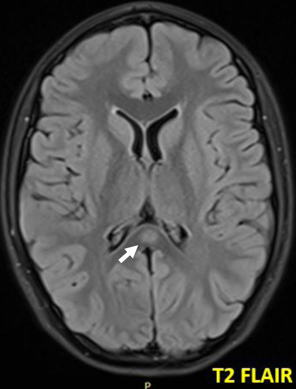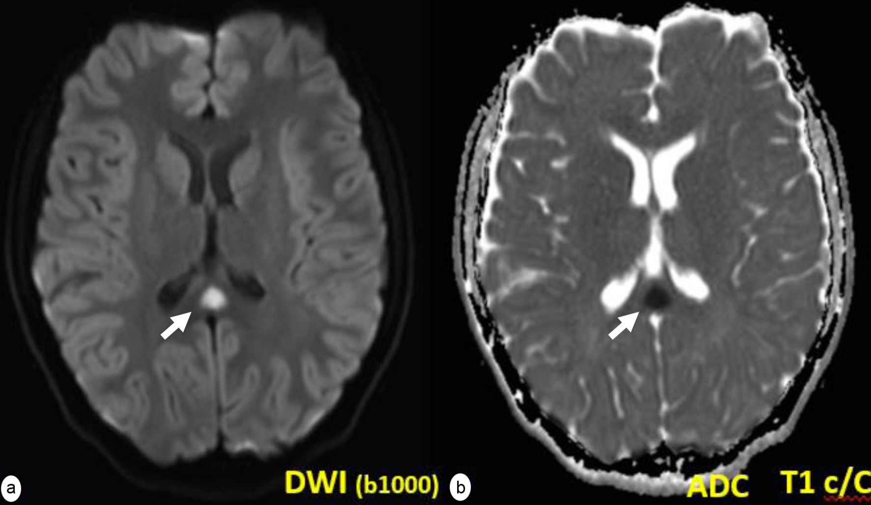
Figure 1. Well-demarcated ovoid lesion (a) in the midline of the splenium of the corpus callosum (white arrows) that does not show uptake after the intravenous administration of paramagnetic contrast (b).
| International Journal of Clinical Pediatrics, ISSN 1927-1255 print, 1927-1263 online, Open Access |
| Article copyright, the authors; Journal compilation copyright, Int J Clin Pediatr and Elmer Press Inc |
| Journal website https://www.theijcp.org |
Case Report
Volume 10, Number 1, March 2021, pages 24-27
Presumed Encephalitis With a Reversible Splenial Lesion
Figures