International Journal of Clinical Pediatrics, ISSN 1927-1255 print, 1927-1263 online, Open Access
Article copyright, the authors; Journal compilation copyright, Int J Clin Pediatr and Elmer Press Inc
Journal website http://www.theijcp.org

Case Report

Volume 8, Number 2, October 2019, pages 37-40


Encephalitis Related to Human Parechovirus Type 3

Figures

Figure 1.
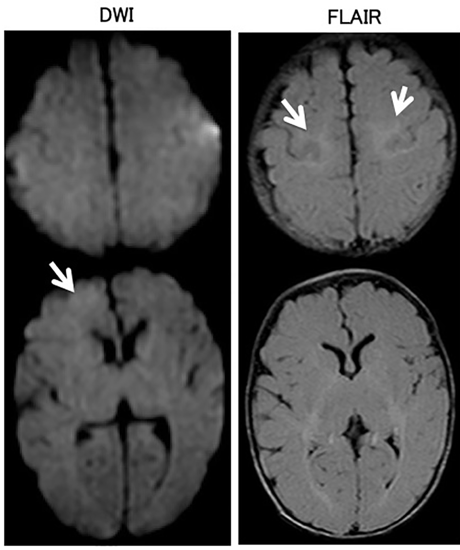
Figure 1. Brain magnetic resonance imaging on the third day of admission. In the brain magnetic resonance diffusion-weighted image (DWI), the right frontal lobe exhibited slightly high-intensity areas. T2-weighted fluid-attenuated inversion recovery (FLAIR) showed hyperintense lesions in bilateral parietal white matter.
Figure 2.
Figure 2. Electroencephalogram (EEG) on day 4 of onset. In the EEG, spike waves were independently recognized in the left and right parietal regions. Slow waves were not observed in the background activity.