Figures
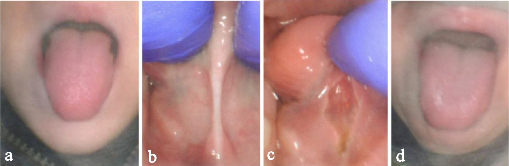
Figure 1. Case 1 of a 5-year-old male with hidden posterior tongue-tie. Before the procedure maximum protrusion (a) and digital elevation (b), immediately after the procedure maximum elevation (c) and maximum protrusion (d).
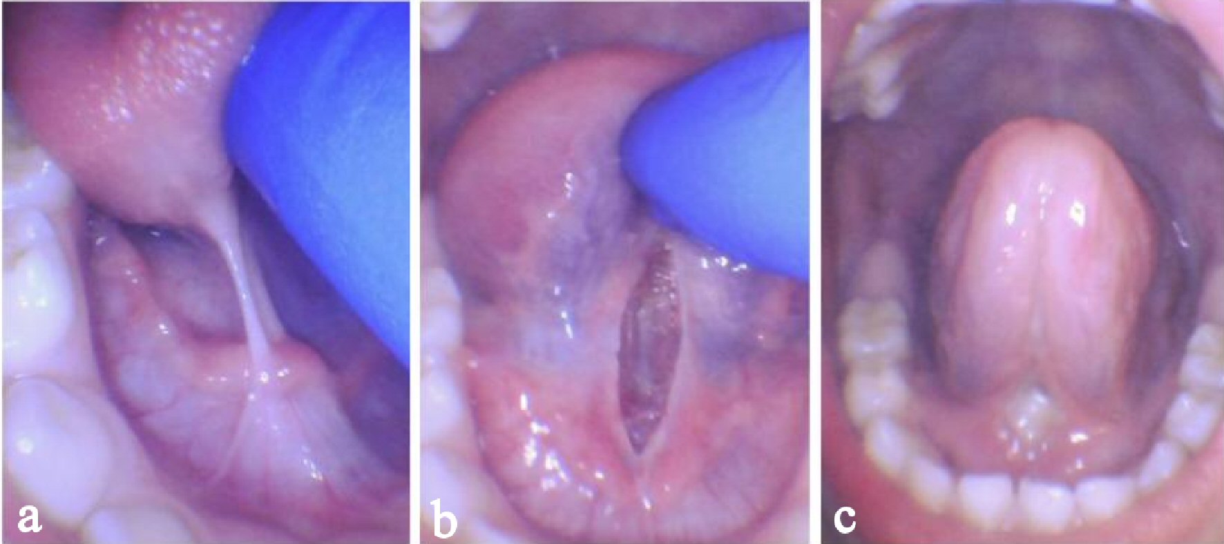
Figure 2. Case 2 of a 5-year-old male with posterior tongue-tie. Before the procedure maximum digital elevation (a), immediately after the procedure maximum digital elevation (b), healing at 1 week showing sustained elevation and increased mobility (c).
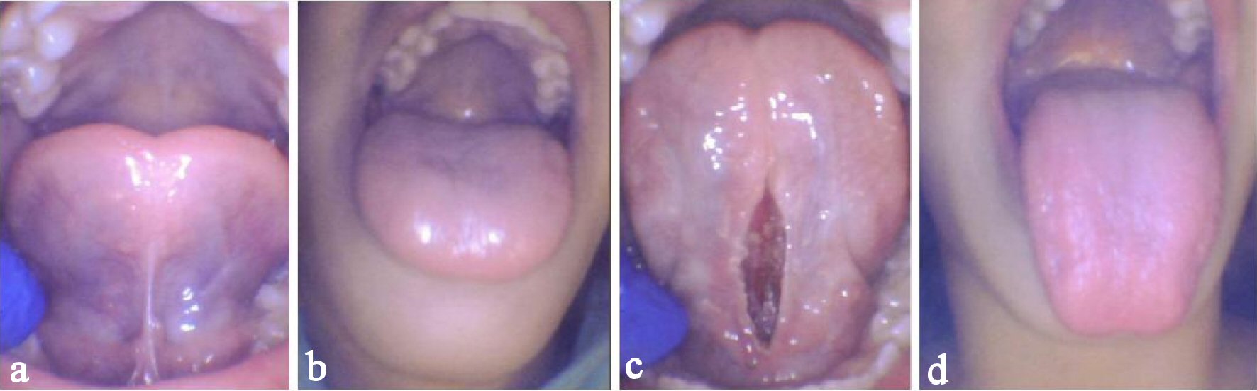
Figure 3. Case 3 of an 11-year-old female with posterior tongue-tie and impaired elevation and protrusion. Before the procedure maximum elevation (a) and protrusion (b), immediately after the procedure maximum elevation (c) and protrusion (d).
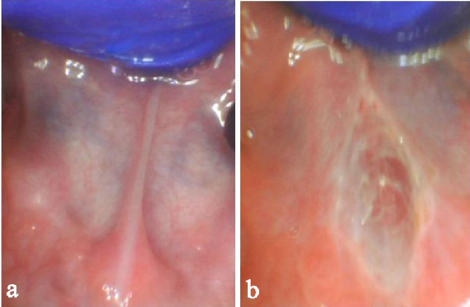
Figure 4. A 2-year-old male with hidden posterior tongue-tie. Before the release maximum elevation (a), immediately after the release elevation demonstrating no bleeding and diamond shaped wound with increased elevation (b).
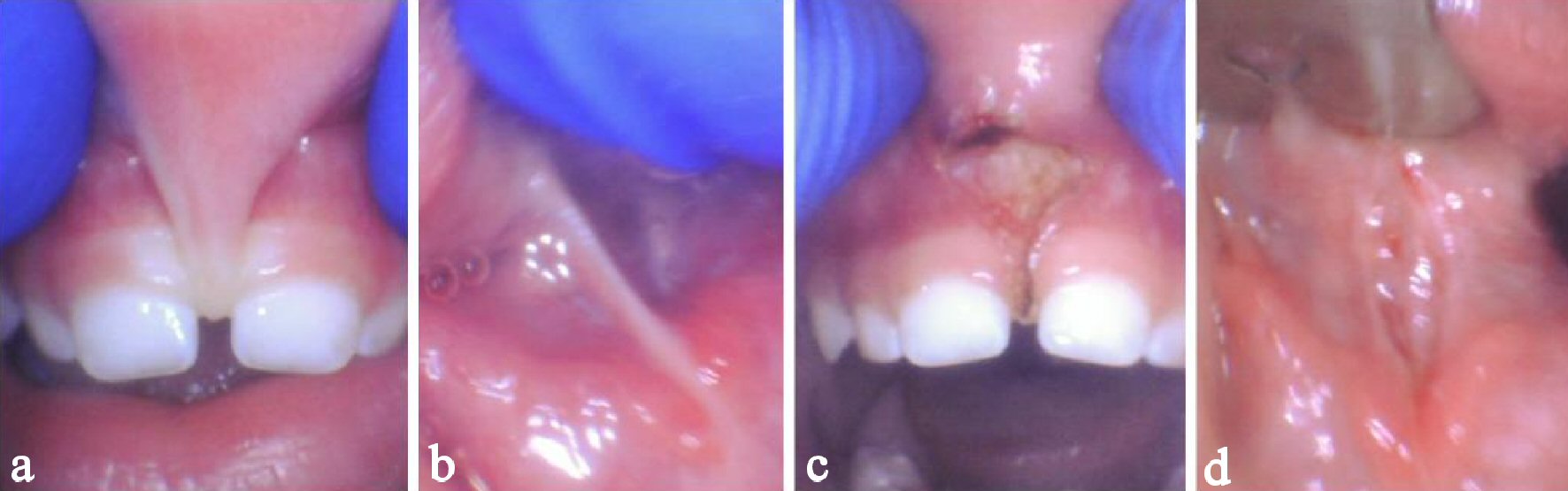
Figure 5. One 17-month-old girl with restrictive maxillary lip-tie and posterior tongue-tie. Before the release lip-tie (a) and posterior tongue-tie (b). Immediately after the release increased maxillary lip elevation (c) and tongue elevation (d).
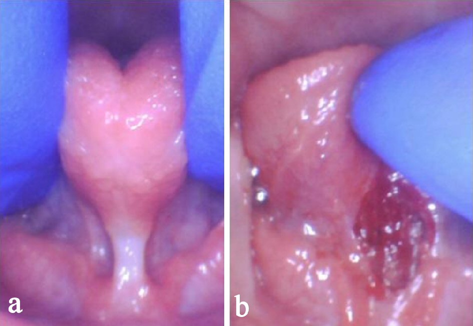
Figure 6. Improperly released tongue after scissors frenotomy showing impaired tongue elevation and thick band of fibrous tissue (a). Immediately after the release showing diamond-shaped wound, hemostasis, and increased elevation of tongue (b).