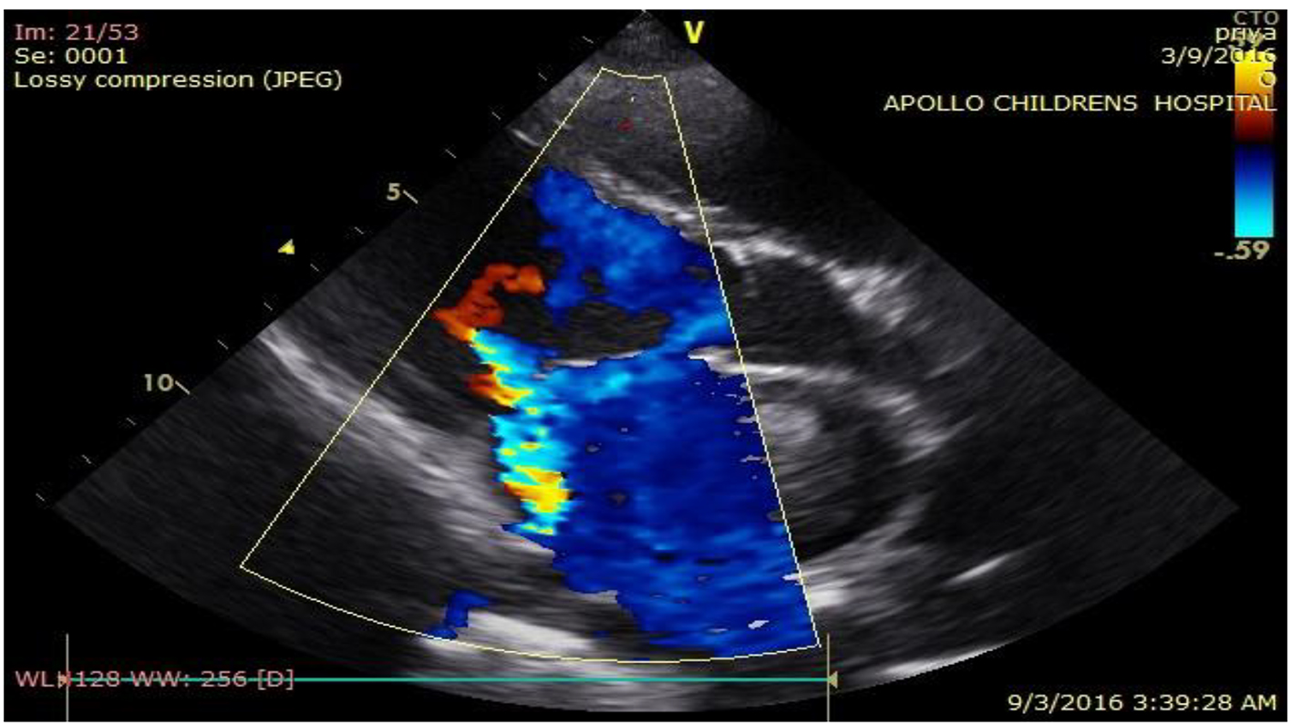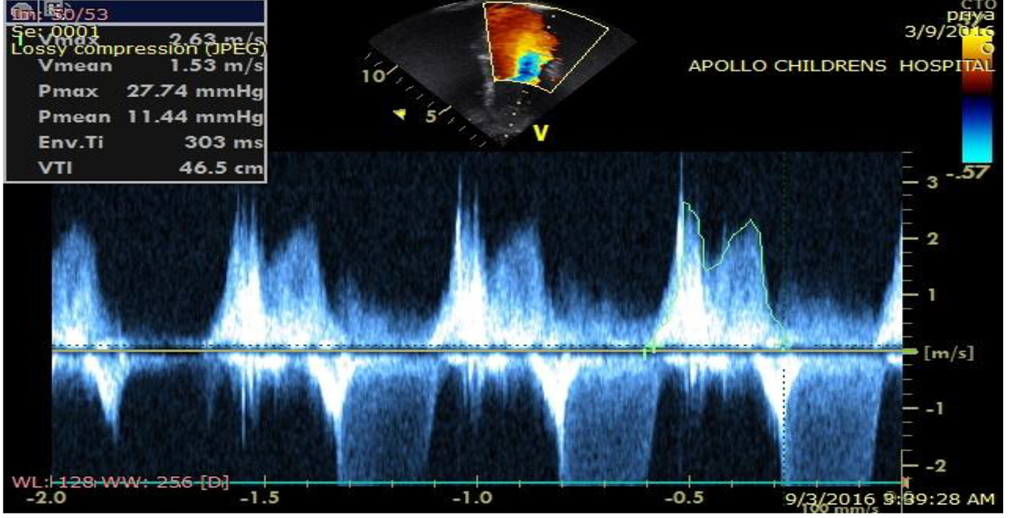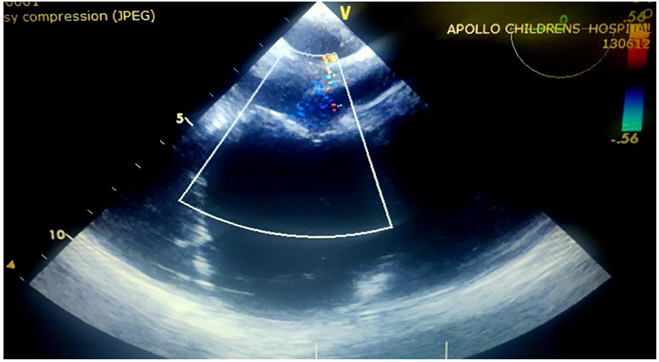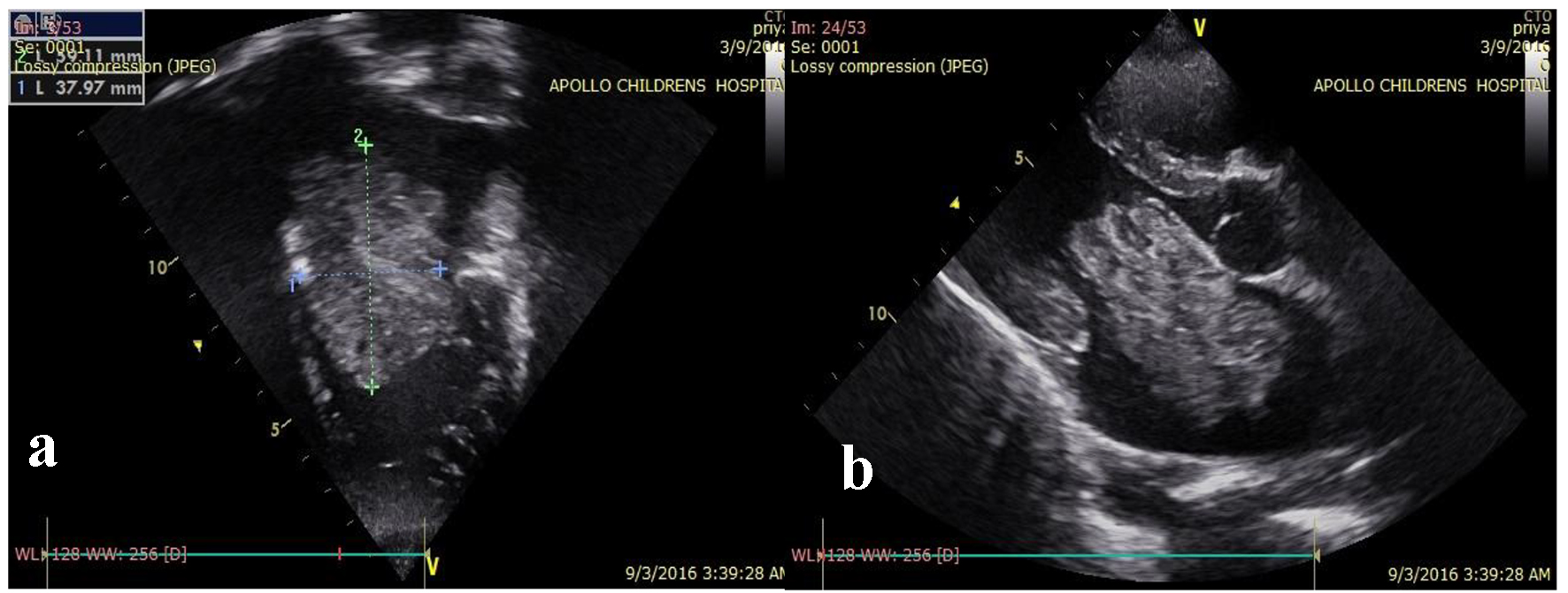
Figure 1. (a, b) Echocardiography showing left atrial mass of homogenous echogenicity, pedunculated, non-cystic, with no calcific focus in four-chamber and parasternal long axis view.
| International Journal of Clinical Pediatrics, ISSN 1927-1255 print, 1927-1263 online, Open Access |
| Article copyright, the authors; Journal compilation copyright, Int J Clin Pediatr and Elmer Press Inc |
| Journal website http://www.theijcp.org |
Case Report
Volume 6, Number 3-4, December 2017, pages 46-50
An Unusual Presentation of Left Atrial Myxoma in an Adolescent
Figures