International Journal of Clinical Pediatrics, ISSN 1927-1255 print, 1927-1263 online, Open Access
Article copyright, the authors; Journal compilation copyright, Int J Clin Pediatr and Elmer Press Inc
Journal website http://www.theijcp.org

Case Report

Volume 4, Number 4, December 2015, pages 189-192


Pulmonary Artery Aneurysm as a Clue to Behcet’s Disease in a 7-Year-Old Boy

Figures

Figure 1.
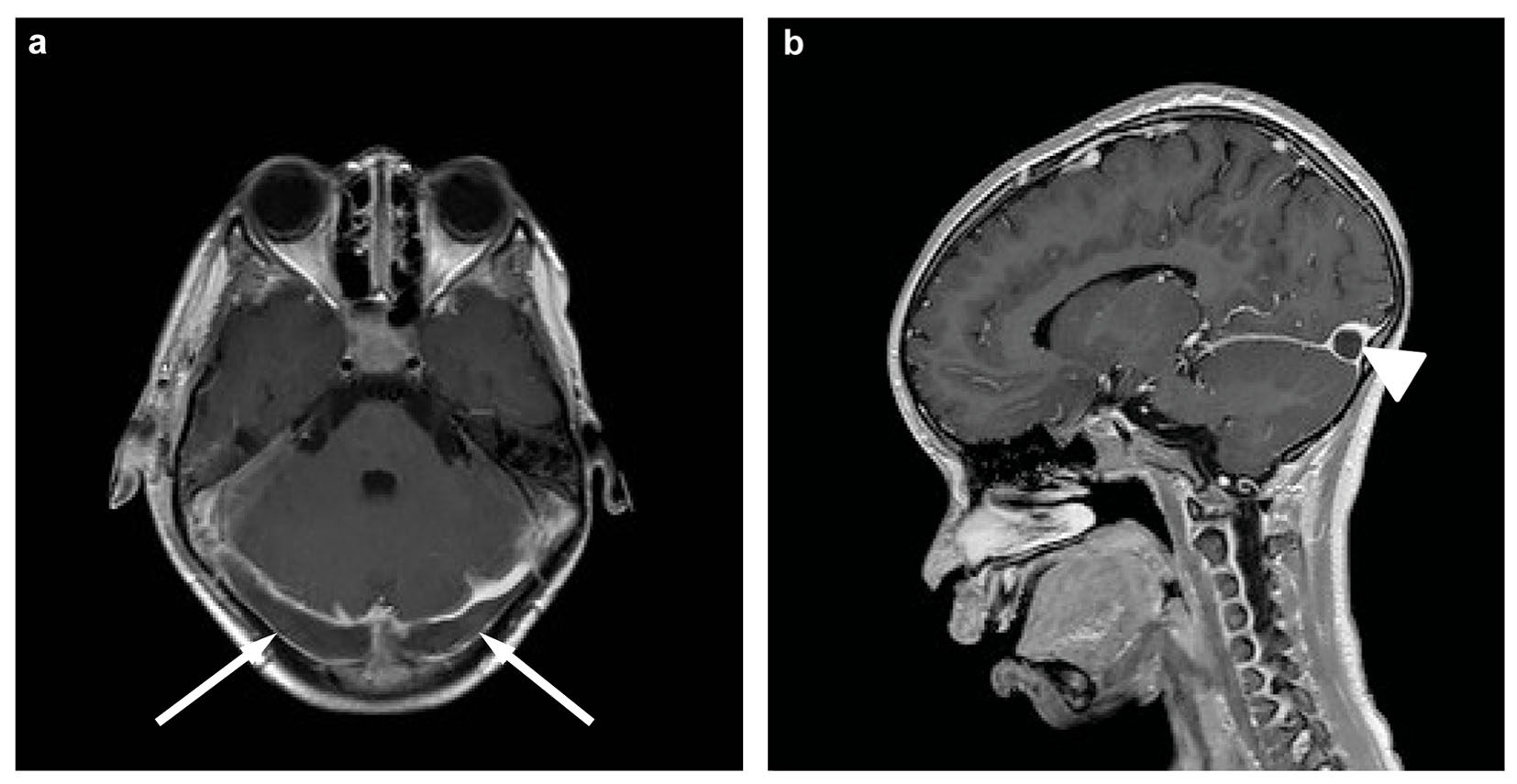
Figure 1. Magnetic resonance imaging of the brain, transverse (a) and sagittal (b) T1-weighted post-contrast. Both images show extensive thrombosis of the transverse sinus on both sides (arrows) and confluens sinuum (arrowhead).
Figure 2.
Figure 2. Contrast-enhanced CT of the thorax: (a) the initial size of the pulmonary artery aneurysm (diameter 1.1 cm), (b) the size of the aneurysm 1 month later (diameter 2.9 cm), also appreciate increase of the mural thrombus.
Figure 3.
Figure 3. Elastica van Gieson stain of (a) pulmonary artery aneurysm (between arrows) with thrombus (T) (× 12 magnification; scale bar = 1,000 µm) and (b) pulmonary artery and inflammatory infiltrate (× 50 magnification; scale bar = 500 µm). There are (1) destruction of lamina elastica of vessel wall and (2) inflammatory infiltrate consisting of macrophages and neutrophilic granulocytes (T: thrombus; B: bronchus; C: cartilage).