International Journal of Clinical Pediatrics, ISSN 1927-1255 print, 1927-1263 online, Open Access
Article copyright, the authors; Journal compilation copyright, Int J Clin Pediatr and Elmer Press Inc
Journal website http://www.theijcp.org

Case Report

Volume 4, Number 2-3, October 2015, pages 162-165


Retroperitoneal Arteriovenous Malformation: A Rare Cause of Pulmonary Hypertension in a Premature Infant

Figures

Figure 1.
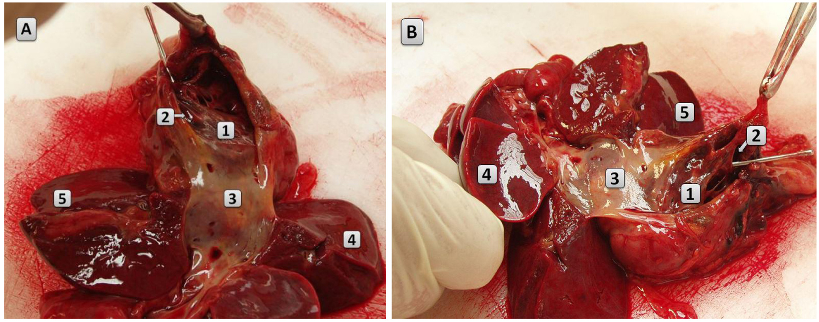
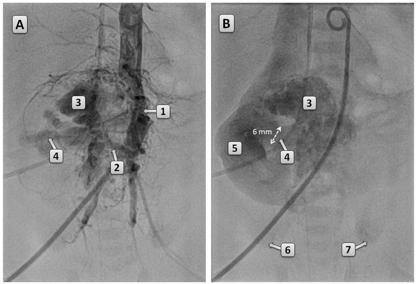
Figure 1. (A, B) DSA of the aorta performed at the age of 18 h. The aorta is dislocated to the left by the AVM. Blood supply of the AVM occurs through the lumbar arteries. In (B), a 6 mm shunt between the AVM and IVC can be seen. Renal perfusion is decreased as there is only slight enhancement at the lower poles of the kidneys. 1: abdominal aorta; 2: feeding vessel of the AVM; 3: AVM; 4: shunt between AVM and the IVC; 5: IVC; 6: lower pole of the right kidney; 7: lower pole of the left kidney.
Figure 2.
Figure 2. (A, B) Macroscopic picture of the AVM. The IVC and the liver are dissected. The AVM is composed of cystic cavities and it encircles most of the suprarenal part of the IVC. 1: AVM; 2: metal probe is inserted in the shunt connecting the AVM and the aorta; 3: IVC; 4: right lobe of the liver; 5: left lobe of the liver.