![Figure 1.](/tables/ijcp149w-g001.jpg)
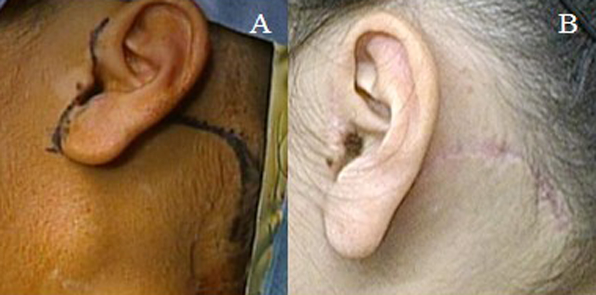
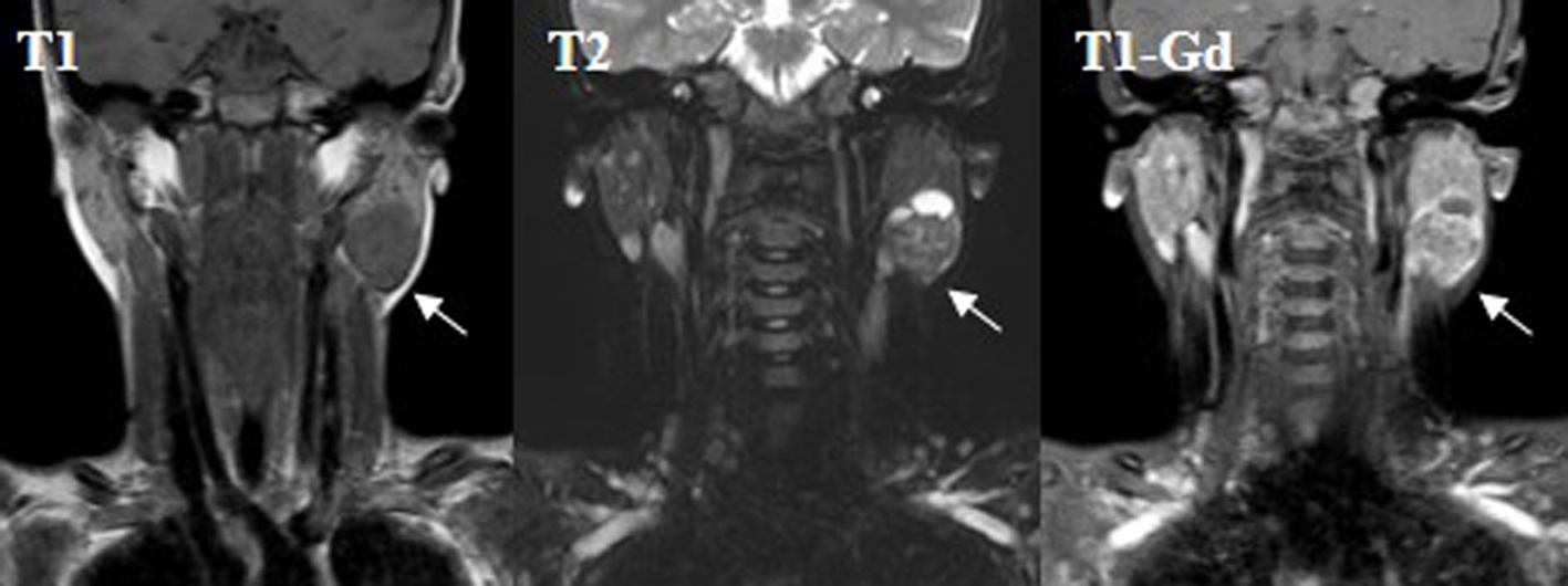
Figure 1. Magnetic resonance imaging (MRI) examination revealed a round mass presenting both cystic and solid lesions within the superficial lobe of the left parotid gland. T1 MRI showed a low signal mass (T1) and T2 MRI showed a relative low signal round mass (T2). The mass with clear margins was slightly enhanced by gadolinium (T1-Gd). The arrows show the tumor mass.
![Figure 3.](/tables/ijcp149w-g003.jpg)
Figure 3. The histopathological findings revealed well encapsulated lesions with cystic and solid areas consisting of an eosiophilic oncocytic epithelial cell component arranged in double layers (A: examination on low power). A variable amount of lymphoid tissue represented with germinal centers (B: examination on high power).