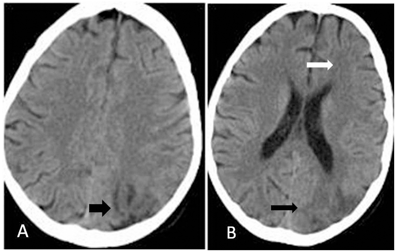
Figure 1. (A, B) Axial CT scan of brain showed effacement of sulci in the left cerebral hemisphere (white arrow) and hypodense region in the subcortical and deep white matter of the occipitoparietal lobe (black arrow).
| International Journal of Clinical Pediatrics, ISSN 1927-1255 print, 1927-1263 online, Open Access |
| Article copyright, the authors; Journal compilation copyright, Int J Clin Pediatr and Elmer Press Inc |
| Journal website http://www.theijcp.org |
Case Report
Volume 3, Number 1, March 2014, pages 29-32
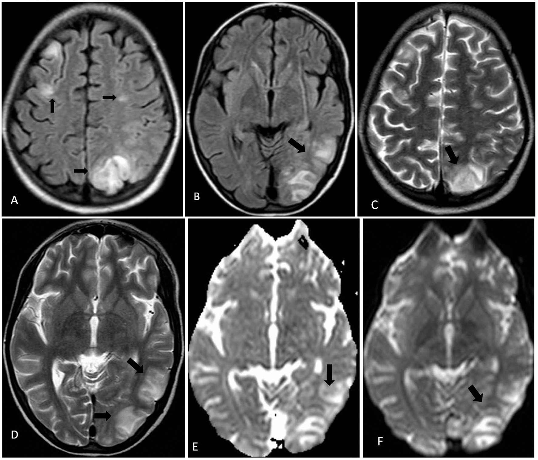
Atypical Imaging Findings of Posterior Reversible Encephalopathy Syndrome in a Child: Case Report and Review of Literature
Figures